
Posterior Urethral
What Is a Posterior Urethral Valve?
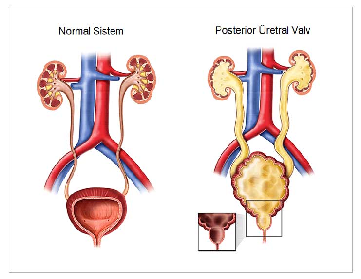
Posterior urethral valves are the most common congenital anomaly causing bladder outlet obstruction. They are observed in approximately one out of every 5,000 to 6,000 male newborns. Obstruction (narrowing) caused by membrane-like folds in the posterior urethra (urinary tract) is observed exclusively in male newborns. This condition can lead to life-threatening emergencies and may also negatively affect kidney and bladder development in the long term.
What Are the Symptoms of a Posterior Urethral Valve?
With the widespread use of ultrasound in prenatal care, the likelihood of detecting these conditions in utero has increased. A distended, thick-walled bladder and bilateral hydroureteronephrosis (swelling of the kidneys and ureters) are among the key findings. Ultrasound evaluation of amniotic fluid is an important indicator. A decrease in amniotic fluid, known as oligohydramnios, is a common finding in patients with posterior urethral valve disease
. Infants with oligohydramnios may experience respiratory distress, which can sometimes be life-threatening. This may be associated with kidney failure. In some cases, valvular bladder syndrome may be observed, which can cause abnormalities in voiding dynamics and lead to problems with urine storage in the bladder. In some cases, vesicoureteral reflux, non-functioning kidneys, and bladder diverticula may be observed.
How Is a Posterior Urethral Valve Diagnosed?
Although the diagnosis of a valve is largely confirmed by the presence of a wide, thick-walled, trabeculated bladder on a postpartum voiding cystourethrogram, the gold standard diagnostic method is cystoscopy (examination of the bladder by inserting a camera through the urinary tract). During cystoscopy, valves can be observed causing a narrowing following an elongated and dilated posterior urethra.
How Is Posterior Urethral Valve Treatment Performed?
⦁ The first step in treatment is to address acute problems. These include respiratory distress, sepsis, and fluid and electrolyte loss.
⦁ Temporary drainage of urine is achieved using a catheter.
⦁ In primary valve ablation, the valve is incised using cold or hot blades with the aid of a thin cystoscope inserted through the infant’s urethra (urinary tract).
⦁ In some cases—especially if the infant is very small or premature—a cystostomy, in which the bladder is temporarily connected to the skin, allows urine to drain freely into a diaper.
⦁ In some cases, a skin stoma may also be created on the ureter as a temporary measure.
⦁ Medical treatment may include emptying the bladder through frequent and regular urination, clean-intermittent catheterization, anticholinergic medications, and antibiotic prophylaxis when indicated.
What Problems Can a Posterior Urethral Valve Cause in the Future?
Due to permanent abnormalities that may cause long-term problems with bladder emptying, frequent infections and kidney damage may occur. This condition often manifests as a decrease in urine concentration and polyuria (increased urine output). In approximately one-third of cases, urinary incontinence occurs due to impaired bladder dynamics.
What Are the Long-Term Goals of Treatment for Posterior Urethral Valve?
⦁ A bladder capable of adequately storing low-pressure urine
⦁ Preservation of renal function
⦁ Prevention of infection
⦁ Maintenance of urinary continence