Flexible URS-RIRS in the Treatment of Kidney Stones
- What Is a Stone?
- How Does It Form?
- Who Is at Risk?
- Other Symptoms Associated with Renal Colic
- Diagnosis
- Treatment
- Conservative Stone Treatment
- Medical Expulsive Therapy
- Active Stone Therapy
- Stone Fragmentation Using Shock Waves (SWL)
- Ureteroscopy (URS)
- Minimally Invasive Kidney Stone Surgery
- Preventing the Recurrence of Stone Formation
What Is a Stone?
A stone is a hard, solid mass
that can form in the gallbladder, bladder, and kidneys. The causes of these types of stones vary, and they are treated in different ways
.
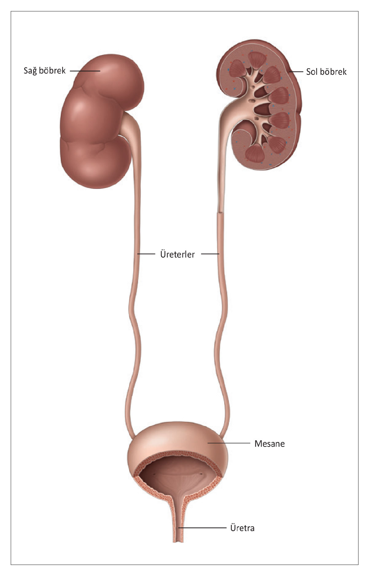
Figure 1
How Does It Form?
Kidney stones form when minerals or acid salts in your urine crystallize. Most stones pass out of your body when you urinate. However, in some cases, you may need treatment to remove the stone.
Who Is at Risk?
Anyone—whether male or female—can develop kidney stones at some point in their life. Kidney stones can form if there is an imbalance in the way your body produces urine. This may be related to the amount of fluid you drink and whether substances that trigger stone formation are present in your urine.
Symptoms
People generally associate kidney and ureteral stones with pain.
However, symptoms vary depending on characteristics such as the stone’s size, shape, and location in the urinary tract
and can range from completely
painless to extremely painful.
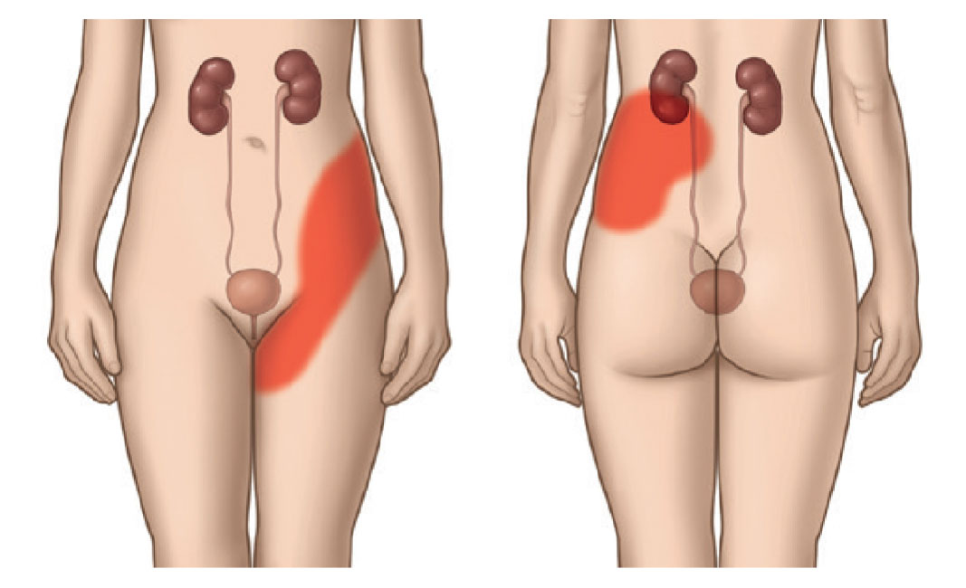
Figure 2
Severe pain (renal colic)
If a stone blocks the normal flow of urine through the ureters, you will experience severe pain known as renal
colic. It is a sharp pain felt in the lower back and flank
(the area on both sides of your body extending from the lower part of the ribs
to the hip). You may also feel the pain in your groin or thigh.
Men may also feel pain in their testicles
Other Symptoms Associated with Renal Colic:
• Nausea
• Vomiting
• Blood in the urine (pink-colored urine)
• Pain when urinating
• Fever
Renal colic is a medical emergency, and you should contact your family doctor or go to the nearest hospital to relieve the pain.
Diagnosis
Your doctor will perform a series of tests to determine what is causing your symptoms
. This is called diagnosis.
To locate your stone, your doctor will need to visualize your internal organs
. To do this, you will need to undergo an ultrasound (also known as an ultrasonogram
), which uses high-frequency sound waves to create images
. In addition to an ultrasound, you may also need an X-ray of your urinary tract
.
Another commonly used diagnostic method is a CT scan
(computed tomography). With this scan, the size, shape, and
thickness of your stone can be clearly seen.
Treatment
Not all stones require treatment. You may need treatment if your stone is causing discomfort and is not passing naturally in your urine. If you have pre-existing medical conditions, your doctor may also recommend treatment.
If you have a kidney or ureteral stone that is not causing any discomfort, treatment is generally not necessary. Your doctor will provide you with a schedule for regular checkups to ensure your condition does not worsen.
Conservative Stone Treatment
If your stone is likely to pass in your urine, your doctor may prescribe medications to make the process easier. This is called conservative treatment.
Most kidney or ureteral stones pass out of your body in your urine. However, depending on the stone’s size and location, it may take some time to pass. When the stone moves, you may experience renal colic (side pain).
Keep this in mind:
• The closer a stone is to the bladder, the greater the likelihood that it will be passed
• The larger the stone, the lower the chance that it will be passed
Medical Expulsive Therapy
Your doctor may prescribe medications to help you pass the stone more quickly and reduce your pain as it moves. This is called Medical Expulsive Therapy (MET) and is most effective for ureteral stones.
During MET, you’ll need to have regular checkups with your doctor at the frequency they recommend. Your doctor will need to check whether the stone is moving and whether your kidney is functioning properly.
Active Stone Therapy
Kidney or ureteral stones should be treated if they cause symptoms. There are three common methods for removing stones: shock wave lithotripsy (SWL), ureteroscopy (URS), and percutaneous nephrolithotomy (PNL).
Which active treatment method is best for you depends on many factors. The most important factor is the symptoms caused by the stone.
Your doctor may recommend different treatment options depending on whether your stone is located in the kidney or the ureter.
Stone Fragmentation Using Shock Waves (SWL)
SWL is performed using a machine that breaks up stones from outside the body. To break up the stone, focused shock waves (short-pulse, high-energy sound waves) are transmitted through the skin to the stone. The stone absorbs the energy of the shock waves, causing it to break into small pieces. The stone fragments are then passed in the urine over the following days or weeks.
Ureteroscopy (URS)
URS is a type of treatment performed using a small-diameter endoscope. URS is common and has a high success rate with a low risk of complications. URS is performed under general or local anesthesia. While you are under anesthesia, your doctor inserts the endoscope through the urethra into the bladder without making any incisions in your body. The stone is removed using a special “basket.”
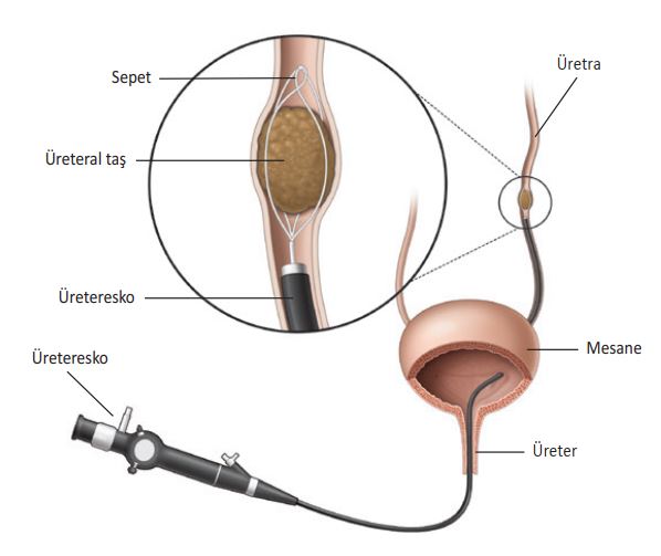
Figure 4
Retrograde intrarenal surgery (RIRS)
Minimally Invasive Kidney Stone Surgery (Percutaneous Nephrolithotomy)
PNL is a surgical procedure in which large stones are removed directly from your kidney.
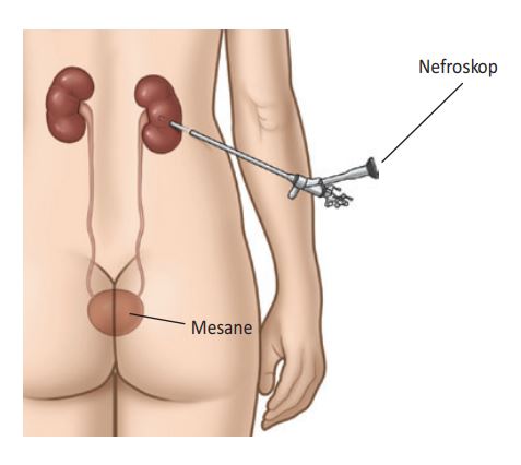
Figure 5-A
The advantage is that even very large stones can be removed in a single procedure. PNL is performed under general anesthesia.
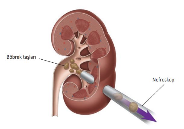
Figure 5-B
Preventing the Recurrence of Stone Formation
Some patients with stones in their kidneys or ureters may develop more stones in the future.
After your stone has passed or been removed through treatment, your doctor will determine whether you are at high risk for stone recurrence. To do this, your doctor will need to analyze your stone. Your doctor will also evaluate the results of the blood and urine tests performed before treatment.
If your risk of stone recurrence is low, general lifestyle changes will be sufficient to reduce the risk of another stone forming.
Recommendations for Adults.
Drink more fluids
• Drink between 2.5 and 3 liters of fluid every day
• Spread your fluid intake evenly throughout the day
• Choose beverages with a neutral pH, such as water or milk
• Keep track of your urine output. It should be 2–2.5 liters per day.
• Check the color of your urine. It should be light in color.
• If you live in a hot climate or engage in intense physical activity, drink more fluids. This will help you replenish lost fluids.
Adjust Your Diet
Depending on your individual circumstances, your doctor may offer recommendations for adjusting your diet. It is important to discuss this with your doctor first.
• Follow a balanced and varied diet.
• Eat plenty of vegetables, high-fiber foods, and fruits (especially citrus fruits).
• Try to eat more foods that are low in oxalates, such as eggs, lentils, white rice, peeled apples, grapes, cauliflower, and zucchini.
• Make sure your diet includes enough calcium (about 1,000 milligrams a day). However, be cautious about calcium supplements and follow the advice of your doctor or nurse.
• Reduce the amount of salt in your diet (it should not exceed 3–5 grams per day)
• Animal protein, which is particularly abundant in the meat of young animals
Don't eat too much of it. Instead, get your protein from vegetables like avocados, cauliflower, and peas.
• Maintain a healthy body weight (Your Body Mass Index should be between 18 and 25 kg/m²)
Healthy Habits
Adopting a healthy lifestyle is always a good idea.
• Try to exercise 2 or 3 times a week
• Avoid stress
What Is a Stone?
A stone is a hard, solid mass that can form in the gallbladder, bladder, and kidneys. The causes of these types of stones vary, and they are treated in different ways.How Does It Form?
Kidney stones form when minerals or acid salts in your urine crystallize. Most stones pass out of your body when you urinate. However, in some cases, you may need treatment to remove the stone. (Figure 1).Who Is at Risk?
Anyone—whether male or female—can develop kidney stones at some point in their life. Kidney stones can form if there is an imbalance in the way your body produces urine. This may be related to the amount of fluid you drink and whether substances that trigger stone formation are present in your urine.Symptoms
People generally associate kidney and ureteral stones with pain. However, symptoms can vary depending on the stone’s characteristics—such as its size, shape, and location in the urinary tract—ranging from completely painless to extremely painful. Severe pain (renal colic) If a stone blocks the normal flow of urine through the ureters, you will experience severe pain known as renal colic. It is a sharp pain felt in your lower back and flanks (the area on either side of your body extending from the lower ribs to the hips). You may also feel the pain in your groin or thigh. Men may also feel pain in their testicles (Figure 2)Other Symptoms Associated with Renal Colic:
• Nausea • Vomiting • Blood in the urine (pink-colored urine) (see Figure 2) • Pain when urinating • FeverRenal colic is a medical emergency, and you should contact your family doctor or go to the nearest hospital to relieve the pain.Diagnosis
Your doctor will perform a series of tests to determine what is causing your symptoms. This is called diagnosis. To locate your stone, your doctor will need to visualize your internal organs. To do this, you will need to undergo an ultrasound (also known as ultrasonography), which uses high-frequency sound waves to create images. In addition to an ultrasound, you may also need an X-ray of your urinary tract. Another commonly used diagnostic method is a CT scan (computed tomography). This scan clearly shows the size, shape, and thickness of your stone.Treatment
Not all stones require treatment. You may need treatment if your stone is causing discomfort and is not passing naturally in your urine. If you have pre-existing medical conditions, your doctor may also recommend treatment. If you have a kidney or ureteral stone that isn’t causing any symptoms, treatment is generally not necessary. Your doctor will provide you with a schedule for regular checkups to ensure your condition does not worsen.Conservative Stone Treatment
If it is likely that your stone will pass in your urine, your doctor may prescribe medications to make this process easier. This is called conservative treatment. Most kidney or ureteral stones pass out of your body in your urine. However, depending on the stone’s size and location, it may take some time to pass. You may experience renal colic when the stone moves.Keep this in mind:• The closer the stone is to the bladder, the greater the likelihood that it will be passed. • The larger the stone, the lower the chance that it will be passed.Medical Expulsive Therapy
Your doctor may prescribe medications (known as alpha-blockers and nifedipine) to help you pass the stone more quickly and reduce your pain as it moves. This is called Medical Expulsive Therapy (MET) and is most effective for ureteral stones. During MET, you’ll need to have regular checkups with your doctor at the frequency they recommend. Your doctor will need to check whether the stone is moving and whether your kidney is functioning properly.Active Stone Therapy
Kidney or ureteral stones should be treated if they cause symptoms. There are three common methods for removing stones: shock wave lithotripsy (SWL), ureteroscopy (URS), and percutaneous nephrolithotomy (PNL). Which active treatment method is best for you depends on many factors. The most important factor is the symptoms caused by the stone. Your doctor may recommend different treatment options depending on whether your stone is located in the kidney or the ureter.Stone Fragmentation Using Shock Waves (SWL)
SWL is performed using a machine that breaks up stones from outside the body. The stone’s To break it up, focused shock waves (short-pulse, high- (energetic sound waves) are transmitted from the skin to the stone. The stone, shockIt absorbs the energy of the waves and thus breaks them down into smaller pieces. The stone particles then remain for the following days oris excreted in the urine within weeks ((Figure 3)Ureteroscopy (URS)
URS is a type of treatment performed using a small-diameter endoscope. URS is widely used and has a high success rate, while the risk of complications is dIt's cold. URS is performed under general or local anesthesia. You will be under anesthesia While you are under anesthesia, your doctor will perform the procedure without making any incisions in your body It enters the bladder through the urethra using an endoscope. The stone is retrieved using a special “basket” is pulled out using (Figure 4).Minimally Invasive Kidney Stone Surgery (Percutaneous Nephrolithotomy)
PNL is a procedure in which large stones are removed directly from your kidneyIt is a surgical procedure. The advantage is that even very large stones can be removed in a single operation is that it can be removed (Figures 5a and b). PNL is performed under general anesthesiais carried out.Preventing the Recurrence of Stone Formation
Some patients with stones in their kidneys or ureters may develop more stones in the future. After your stone has passed or been removed through treatment, your doctor will determine whether you are at high risk for stone recurrence. To do this, your doctor will need to analyze your stone. Your doctor will also evaluate the results of the blood and urine tests performed before treatment.If your risk of stone recurrence is low, general lifestyle changes will be sufficient to reduce the risk of developing another stone.Below are some recommendations for adults.
Drink More Fluids• Drink between 2.5 and 3 liters of fluid every day• Spread your fluid intake evenly throughout the day• Choose beverages with a neutral pH, such as water or milk• Monitor your urine output. It should be 2–2.5 liters per day• Check the color of your urine; it should be light in color.• If you live in a hot climate or engage in intense physical exercise, drink more fluids. This will help compensate for fluid loss.Adjust Your Diet
Depending on your individual circumstances, your doctor may recommend adjustments to your diet. It is important to discuss this with your doctor first.• Follow a balanced and varied diet.• Eat plenty of vegetables, fiber-rich foods, and fruits (especially citrus fruits).• Try to eat more foods that are low in oxalates, such as eggs, lentils, white rice, peeled apples, grapes, cauliflower, and zucchini.• Make sure your diet contains enough calcium (about 1,000 milligrams per day). However, be careful with calcium supplements and follow the advice of your doctor or nurse.• Reduce the amount of salt in your diet (it should not exceed 3–5 grams per day).• Do not consume too much animal protein,which is abundant especially in young animal meat. Instead, get your protein from plant sources such as avocados, cauliflower, and peas.• Maintain a healthy body weight (your Body Mass Index should be between 18 and 25 kg/m²).Healthy Habits
Adopting a healthy lifestyle is always a good idea.• Try to exercise 2 or 3 times a week• Avoid stressSend Your Message