Prostate MRI Fusion Biopsy
How is prostate cancer diagnosed?
Every man should undergo a prostate exam and have his serum PSA level checked once a year starting at age 50. For patients with a family history of prostate cancer, these screenings should begin at age 45. Suspicion of prostate cancer usually arises when PSA levels in the blood are elevated and/or suspicious findings are detected during a prostate exam. Patients with suspected prostate cancer should be recommended to undergo a biopsy.
What Do We Learn from a Prostate Biopsy?
The primary purpose of a biopsy is to determine whether a patient has cancer. In addition, the characteristics of the cancer are evaluated, and a treatment approach can be determined based on this information.
Why is fusion biopsy with multiparametric MRI important?
Prostate cancer is the most common cancer in men, and the standard diagnostic method involves a systematic prostate biopsy of 10–14 samples guided by transrectal ultrasound (TRUS). With this method, cancer detection rates range from 27% to 40%; 20% to 25% of clinically significant cancers are missed, and a significant number of clinically insignificant cancers are detected. Patients in whom clinically significant cancer is not detected may not receive the necessary treatment, and a second or even third biopsy may be required.
Multiparametric magnetic resonance imaging, a technique that has been developed and widely adopted in recent years, allows for a detailed examination of the prostate. It is possible to determine whether cancer is present in the prostate, and if so, its location, size, and—perhaps most importantly—whether the cancer is aggressive. Even cancerous lesions as small as 2–3 mm can be detected using this method. For this reason, using this method prior to a biopsy in patients with clinically suspected prostate cancer will ensure that the indication for biopsy is correctly established and will protect our patients from unnecessary biopsies. Therefore, it is extremely important to use multiparametric MRI prior to a biopsy and, if suspicious lesions are identified during this examination, to utilize MRI guidance during the biopsy procedure.
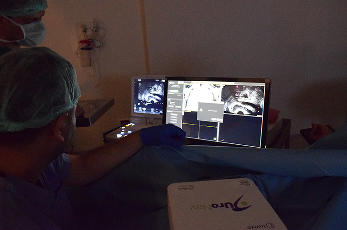
How is an MRI-guided targeted fusion biopsy performed?
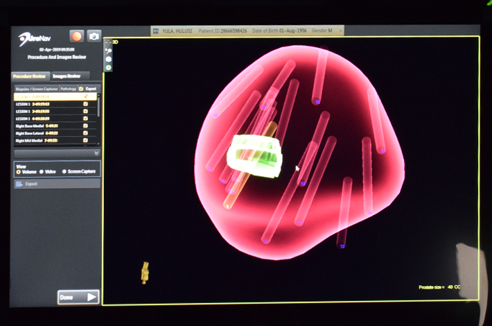
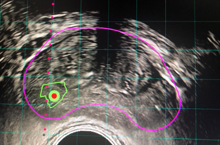
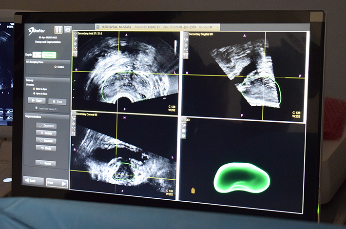
In the targeted prostate fusion biopsy method, multiparametric prostate MRI images are processed using specialized software to create a 3-dimensional model of the prostate. Areas suspected of containing cancer are marked on the 3-dimensional image. These 3-dimensional images are then transferred to the biopsy platform. During the biopsy, prostate images obtained from the TRUS device are converted into 3-dimensional images on the same device, and the fusion process is then performed by superimposing the MRI and TRUS images. Following this, biopsies are taken from the marked suspicious MRI regions with deviations of only a few millimeters. Thanks to this system, biopsies can be taken directly from the marked areas.
What are the clinical benefits of MRI-guided fusion technology?
- Unlike a standard blind biopsy, multiparametric MRI provides 3-dimensional images, allowing for sampling with minimal deviation and error from areas at risk of containing a tumor.
- Compared to standard biopsies, this method detects 30% more cases of cancer.
- The need for repeat biopsies is reduced.
- The diagnosis of clinically insignificant cancers—that is, cancers that pose no harm to the patient—is significantly reduced through the fusion biopsy method. This, in turn, prevents unnecessary major surgeries from being performed.
- Compared to systemic biopsy, it results in a 30% increase in the detection of clinically significant—that is, aggressive—cancers.
- The biopsy sites are precisely identified and can be recorded. This offers a significant advantage for patients on what we call the "active surveillance" follow-up protocol.
In conclusion;
The MRI-guided fusion biopsy method has revolutionized the diagnosis of prostate cancer by overcoming the limitations of conventional biopsy. Fusion prostate biopsy allows for direct biopsy sampling from areas identified by MRI as suspicious for cancer. It also helps prevent unnecessary treatments by reducing the diagnosis of clinically insignificant disease. At the same time, it is highly effective in detecting active cancers.
In light of this information, it is recommended that all men at risk for prostate cancer follow a diagnostic pathway that includes an MRI before the biopsy and a fusion biopsy afterward.
Send Your Message