Ureteropelvic Junction Obstruction (Renal Outflow Obstruction)
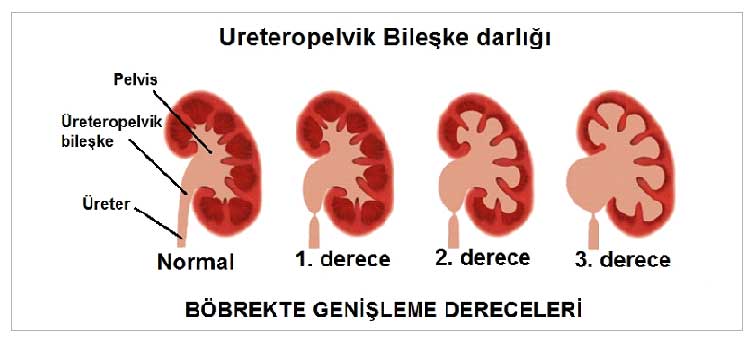
Where Is the Ureteropelvic (UP) Junction?
Urine produced by the kidneys is first collected in a reservoir called the renal pelvis and then transported to the bladder via tube-like structures called ureters. The point where the renal pelvis meets the ureters is called the ureteropelvic junction. A narrowing that develops at this point is defined as ureteropelvic junction obstruction.
The ureteropelvic junction is the site where obstruction (narrowing) is most commonly observed in the upper urinary tract. UP obstruction is present in as many as 64% of children born with hydronephrosis (kidney enlargement).
For urine produced in the kidneys to pass from the renal pelvis into the ureter, a structurally sound ureteropelvic junction is required. In addition, the rhythmic contractions of the ureter—known as peristalsis—also aid in this transport. Both of these components must be structurally sound to ensure proper urine flow. Problems at the ureteropelvic junction can be intrinsic to the ureter itself or extrinsic in origin. Narrowing of that section of the ureter is the most common cause. In addition, there may be extrinsic problems, such as abnormally positioned blood vessels.
What Are the Symptoms of Ureteropelvic Junction Obstruction?
Because urine cannot be excreted from the kidneys—or can only be excreted in very small amounts—it accumulates in the kidneys. Over time, this accumulated urine can cause the kidneys to swell. Kidney swelling (hydronephrosis) is one of the most common symptoms of upper urinary tract (UP) obstruction.
⦁ In some patients, abdominal swelling may be visible from the outside.
⦁ Increased pressure in a kidney that has swollen due to urine buildup can cause pain. Severe pain, particularly in the groin, is a common symptom of a Ureteropelvic junction (UPJ) obstruction.
⦁ Blood in the urine, changes in urine color, inflammation, and related infectious diseases can also be considered symptoms of UP stricture
⦁ Although these symptoms may not be clearly identifiable in children, it is important to keep in mind that conditions such as restlessness, recurrent urinary tract infections, and decreased urine output may indicate an upper urinary tract (UP) stricture
.
Individuals experiencing one or more of these symptoms at the same time should consult a specialist without delay.
How Is Ureteropelvic Junction Stenosis Diagnosed?
Most ureteropelvic junction obstructions are now detected in utero. If not detected in utero, symptoms such as an abdominal mass due to kidney enlargement, blood in the urine, and infection may be observed in the neonatal period. In older children, abdominal or flank pain, blood in the urine—especially following minor trauma—and infection may be observed. It is noteworthy that flank pain tends to worsen when urine output increases (e.g., after excessive fluid intake or consumption of caffeinated beverages).
How Is Ureteropelvic Junction Stenosis Monitored?
Patients with hydronephrosis detected in utero should be re-evaluated with ultrasound after birth to determine whether the hydronephrosis persists. However, this procedure should be performed no earlier than 3 days after birth. In severe cases involving bilateral severe hydronephrosis, however, this waiting period may not be feasible.
If an ultrasound performed at the appropriate time after birth confirms dilation of the renal collecting system, a voiding cystourethrogram and renal scintigraphy should be scheduled. If the ultrasound does not show the presence of hydronephrosis after birth, further testing is not necessary.
Although voiding cystourethrography is traditionally recommended for all infants with hydronephrosis, based on the results of recent studies, it is appropriate to limit the use of this examination, which is quite stressful for both the infant and the family. The purpose of this examination is to rule out vesicoureteral reflux and to evaluate the posterior urethra in male infants. The ideal time for this examination is 4 to 6 weeks after birth. However, immediate examination is required in cases of posterior urethral strictures—known as posterior urethral valves—which are specific to male infants. If reflux is not detected during this examination, hydronephrosis is evaluated using a scintigraphic examination called a diuretic renogram. There are two main radioisotopic agents used for this examination: DTPA and MAG3. Both determine total and individual kidney functions. The curves showing the clearance of these radioactive substances from the renal pelvis also provide guidance regarding the presence or degree of obstruction. To ensure the results are not compromised during the examination, placement of a urethral catheter is recommended. The response to the diuretic is also important when analyzing the clearance curves. However, particularly in cases of hydronephrosis detected in utero and evaluated during the neonatal period, it is important to know the percentage contribution of each kidney to total renal function, a concept known as differential renal function. A loss of function of 10% or more in the affected hydronephrotic kidney compared to the contralateral side is considered significant.
How Should Ureteropelvic Junction Stenosis Be Treated?
Ureteropelvic junction obstruction is a treatable condition. The key here is to ensure close monitoring of patients and to establish an appropriate treatment plan. In most cases, symptoms are mild. In such cases, routine follow-up of the patient will suffice. During this routine follow-up, the patient must undergo various tests at regular intervals, such as ultrasounds, blood tests, and urinalysis. This routine follow-up continues until the obstruction or stricture has completely resolved. In some cases, symptoms resolve on their own. In other cases, medication may be sufficient.
Surgical treatment is necessary when ureteropelvic junction (UPJ) obstruction is symptomatic and impairs kidney function. The ureteropelvic junction is removed via laparoscopic or open surgery, and the ureter is re-anastomosed—that is, sutured—to the renal pelvis. If there is a vascular crossing,the ureteropelvic junctionbelow the crossing vessel is repositioned abovethe vessel.
Send Your Message
