Doctor's Office

0322 503 80 70

WhatsApp & Mobile

0543 333 33 34

Comments & Opinions

Patient Testimonials

Testicular Cancer

Why Is the Importance of Testicular Cancers Increasing?

Testicular cancers are more common, particularly in high-income countries.

Among solid tumors—what we refer to as mass tumorstreatment success rates remain high even in cases of metastatic disease (spread to other sites).

Complete remission is possible in 85–90% of patients. However, because these tumors have the potential to spread rapidly, surgery and treatment are required as soon as possible.

What Are the Reasons for Seeking Treatment for Testicular Cancers?

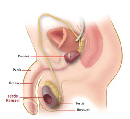
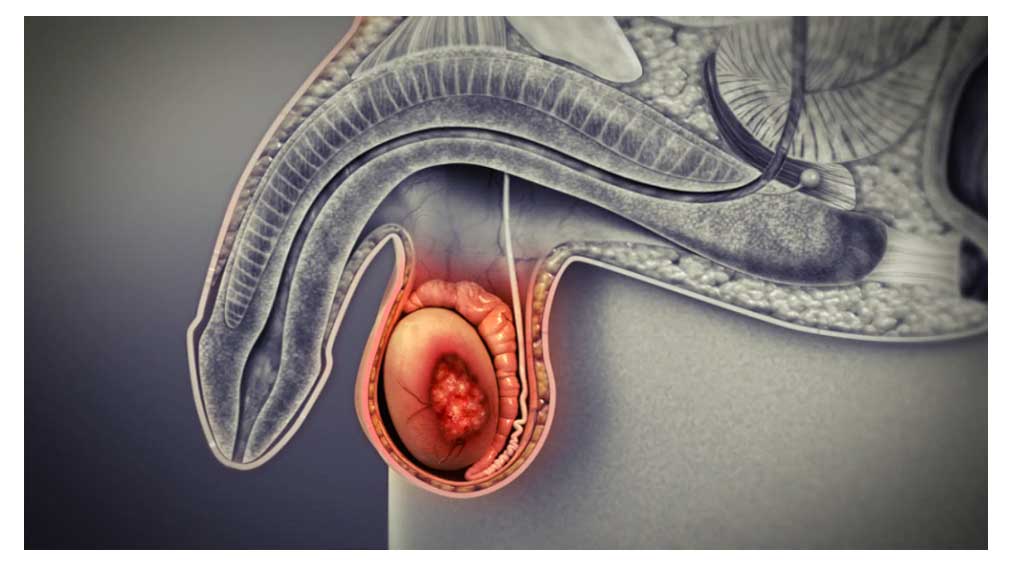
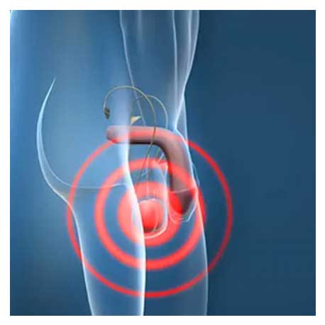
Patients typically present with painless swelling and a mass (hardness) on one side. Twenty percent of patients present with scrotal pain (pain in the scrotum—the skin and subcutaneous tissue covering the testicles). 

Unless proven otherwise, painless scrotal induration should be considered a testicular tumor—unless there is a history of inflammatory conditions—and managed accordingly.

Back and flank pain occur in 11% of cases; in addition, rare symptoms such as hemoptysis (coughing up blood), nausea, vomiting, convulsions (seizures), and bone fractures may occur in 10–20% of cases due to metastatic masses. Gynecomastia (enlarged breasts) may occur in 7% of patients.


How Is Testicular Cancer Diagnosed?

In a suspected case, a physical examination reveals a hard mass in the entire testicle or in a specific area. In such cases, the patient is considered to have testicular cancer until proven otherwise. 

During the examination, the patient is screened for lymph node and organ involvement, as well as breast enlargement, since the condition may have spread throughout the body. It is essential to distinguish between acute epididymo-orchitis (inflammation of the testis and the overlying organ, the epididymis) and testicular cancer; clinically, fever, pain, swelling, skin redness, and increased warmth are indicative of epididymo-orchitis, but a clear distinction may not always be possible. 

The AFP, β-HCG, and LDH tests—known as testicular cancer tumor markers—must be performed. In addition, the diagnosis should be supported by a scrotal ultrasound; ultrasound has a sensitivity of nearly 100% for detecting masses.


How Is a Definitive Diagnosis Made for Testicular Cancer?

A diagnosis can be made with a high degree of certainty through a physical examination, tumor markers, and scrotal ultrasound. Even if the diagnosis is not fully confirmed by these tests, clinical suspicion

In such cases, it is recommended that the affected testicle be surgically removed and examined pathologically. A definitive diagnosis is established following the pathological examination of the removed testicular tissue.

What Should Be Done If a Patient Has Cancer in One Testicle or If Cancer Is Suspected in the Other Testicle at the Same Time?

If the patient has only one testicle, and the mass is not too large and can be removed, it may be possible to attempt to remove only the mass (testicular-sparing surgery). Alternatively, if the patient has tumors in both testicles, testicular-sparing surgery may be performed on at least one of them.
This approach is attempted if the mass is smaller than 2 cm. Patients in this situation require close follow-up after surgery.

What Happens After a Preliminary Diagnosis of Testicular Cancer?

When testicular cancer is suspected, an incision is made in the groin to remove the testicle and the cord—which contains the testicles and the structures surrounding them, including the male glands and blood vessels—as a single unit. This procedure is called a radical inguinal orchiectomy.

 
It is considered a urological emergency requiring surgery. Because testicular cancer has the potential to spread rapidly, the procedure must be performed without delay.

How Is Testicular Cancer Monitored After a Definitive Diagnosis?

Once the disease has been pathologically confirmed, it is essential to determine without delay whether it has spread to other areas. To this end, a full abdominal CT scan, a lung CT scan, and blood tests to measure tumor marker levels should be performed at regular intervals following surgery.

What Is the Postoperative Treatment and Follow-Up for Testicular Cancer?

Treatment and follow-up are determined based on the pathology of the resected tumor and the stage of the disease. Treatment planning is based on the classification of patients according to prognostic factors. In general, for tumors with a good prognosis that have not metastasized and are confined to the testis, follow-up consists of periodic tumor marker testing, physical examinations, and imaging studies. For some patients (stage I seminoma), prophylactic radiation therapy or a single course of chemotherapy may be administered due to the potential for spread to the posterior abdominal wall; alternatively, if the patient is suitable for close follow-up, they may be monitored.

  •  For Stage I non-seminomatous tumors, treatment options may include chemotherapy, retroperitoneal lymph node dissection (removal of lymphatic tissue surrounding the major blood vessels in the posterior abdominal wall), or observation.
  •  Radiation therapy is used to treat stage II disease—seminoma.
  •  For Stage II and higher (metastatic) cases, treatment consists of chemotherapy. Depending on the risk profile, 3 or 4 cycles of chemotherapy are administered. After each cycle of chemotherapy, patients are re-evaluated and undergo either RPLND (retroperitoneal lymph node dissection) or follow-up, as appropriate. When the right chemotherapy is administered at the right time, the cure rates for metastatic testicular cancer are quite high. However, patients may experience side effects from these drugs during chemotherapy; they should be informed of this, and treatment should only begin after obtaining their consent. Patients should be re-evaluated at the end of each cycle (chemotherapy treatment period).

In some patients, adjuvant or secondary chemotherapy is administered when resistance to primary chemotherapy is observed or when residual disease (viable tumor cells) is reported in the postoperative pathology report. A very small group of patients, however, experiences disease progression despite these treatments; for these patients, more intensive (rescue chemotherapy) regimens, rescue surgery, or high-dose chemotherapy in combination with a bone marrow transplant may be administered.

Are There Any Preventive Measures for Testicular Cancer?

Although there is no known preventive method for testicular cancer, treatment success rates are quite high when the disease is detected in its early stages. Men between the ages of 15 and 35—the age group most commonly affected—should be advised and taught to perform a self-examination of their testicles at least once a month (similar to how women perform breast self-exams to screen for breast cancer). Anyone who suspects an abnormality in their testicles should see a doctor without delay.

How Should People with Undescended Testicles Be Evaluated?

In normal development, the testicles descend from the back of the abdomen (at the level of the kidneys) into the scrotum at birth. In some individuals, this process may be delayed, but by around 1 year of age, the testicles have descended in 90% of children. Individuals with undescended testicles should be evaluated, and a decision should be made regarding surgery or medical treatment. For testicles that cannot be brought down into the scrotum—particularly those remaining inside the abdomen—removal (orchiectomy) is recommended.
Men whose testicles descend into the scrotum later than normal should be informed about the risk of testicular cancer.

Send Your Message

    Ali Şahin Design